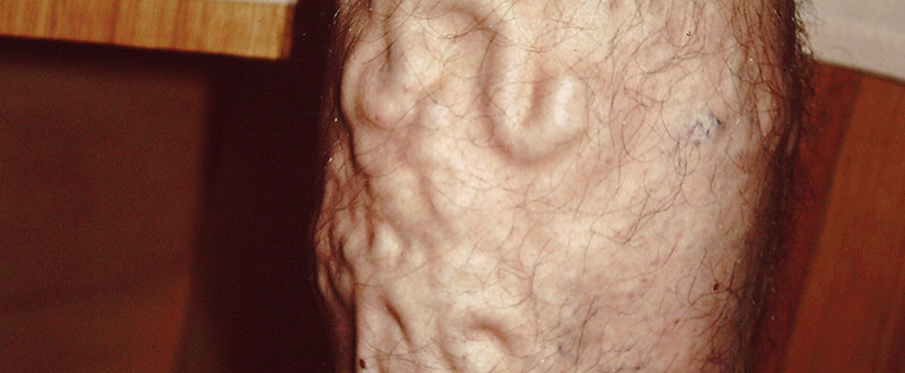
What are Varicose Veins?
The term means ‘enlarged, dilated and tortuous’. Varicose veins can occur in a number of areas on the body but by far the commonest area is in the legs. This is thought to be because standing and walking place extra pressure on leg veins.
Varicose veins are blue in colour because of the thickness of the vein wall and because the blood in these veins has less oxygen.
Are there different types of Varicose veins?
Varicose veins are graded from Grade 1 to Grade 4 depending on how large they are and whether they bulge above the surface level of the skin (with Grade 4 being the largest).
Reticular varicose vein is a term referring to larger blue veins that feed into an area of spider veins .The word Reticular comes from the Latin reticulum meaning ‘little net’ and means relating to or forming a network. Reticular veins feed into areas of spider veins, and hence form a network of veins. When treating spider veins these feeder veins usually also need to be treated for the best results.
Truncal varicose vein is a term referring to a vein that comes from one of the main two vein systems in the legs, either the one on the inside of your leg which is called the Great Saphenous Vein System, or the other on the back of the knee called the Short Saphenous Vein System.
What are the primary causes?
Vein drainage should be from the surface veins inwards to the deep veins and then upwards towards the heart. Varicose veins develop in these superficial vein systems when the one way valves inside these main veins become faulty. If the valves inside these surface veins become stretched and faulty it results in blood pushing back to the surface of the skin and causes the veins to bulge.
It is not clear exactly why vein walls stretch and the valves become faulty in some people and not others. Studies have shown that there is reduced elastin content in the walls of varicose veins compared with normal veins and scientists have also identified proteins that can bind to DNA and start the process that causes varicose veins. It appears there is probably a primary inherited structural issue that predisposes forming of this condition under secondary pressure on the valve and vein walls.

Figure A shows a normal vein with a properly working valve; there is normal blood flow. In Figure B, the varicose vein has a faulty valve, there is abnormal blood flow, the walls of the vein are thin and stretched. The image in the middle shows where in a leg the varicose vein might appear.
Who is at the greatest risk?
Estimates have the incidence of around 30% of adults.
Risk factors that identify a higher risk of developing varicose veins and include:
Sex: Women are much more likely (around twice the incidence)
Genetics: Strong link to a family history
Obesity: Higher incidence in overweight
Pregnancy: Time of increased incidence and worsening severity. Weight gain, pelvic pressure, increased blood volume and hormonal changes all have been implicated.
Age: Increasing incidence as people get older, presumably from the cumulative effect of more years standing and walking on the legs.
Occupation: There is an association with jobs that involve prolonged periods of standing
What are the symptoms?
In the majority of cases there is no significant pain associated, however possible symptoms resulting from congestion include heaviness, tiredness, cramping, aching, pain, and swelling. There is also an increased incidence of restless leg syndrome in patients with varicose veins.
Whilst the greater the degree of venous reflux (backflow) and the larger the varicose veins the more likely the presence of symptoms, it is not a direct correlation. Some patients with very large cases of this condition can be asymptomatic whereas some patients with a fairly small condition can have significant symptoms.
What are the possible complications?
Longstanding back pressure in the veins can result in Chronic Venous Insufficiency (CVI). This can cause skin changes including brownish pigmentation (staining from iron in the blood) and excema (red,dry,itchy skin).
In severe cases it can cause irregular white patches at the ankles (atrophie blanche), shiny, hardened skin (lipodermatosclerosis) or ulceration (Venous Ulcer).
There is an association with an increased risk of developing deep venous thrombosis (DVT) and superficial thrombophlebitis (SVT).
In the presence of venous congestion minor injury to the skin can result in more prolonged bleeding and the skin will also heal more slowly.
What are the treatment options?
Varicose Veins on the surface of the skin can be treated by surface sclerotherapy if there is no significant underlying venous reflux from deeper veins. This may be by liquid or foam sclerotherapy depending on the size of the veins. Surface varicose veins are too large for treatment by surface laser or surface radiofrequency to be effective.
If there is underlying reflux then an Ultrasound guided procedure is required to control the underlying reflux. A Mapping scan will determine which of the Ultrasound procedures is optimal. The choice of Ultrasound Guided treatment is between Ultrasound Guided Sclerotherapy (UGS), Ultrasound Guided laser (also called Endo Venous Laser Therapy) or Ultrasound Guided Radiofrequency (also called VENEFIT).
What if I have both Spider and Varicose Veins?
It is quite common for many patients to have both of these conditions. If these conditions are present are in the same area as a general principle the larger and deeper varicose veins are treated first. Treating these first does not usually result in any significant improvement in the spider veins and they will need to be treated as a secondary procedure. If spider veins are treated first without controlling the underlying deeper vein reflux the results are not as effective and also the chance of getting side effects such as pigmentation and bruising are greater.